Pro and Con: How Important Is the Exact Location of Adductor Canal and Femoral Triangle Blocks?

Highlights
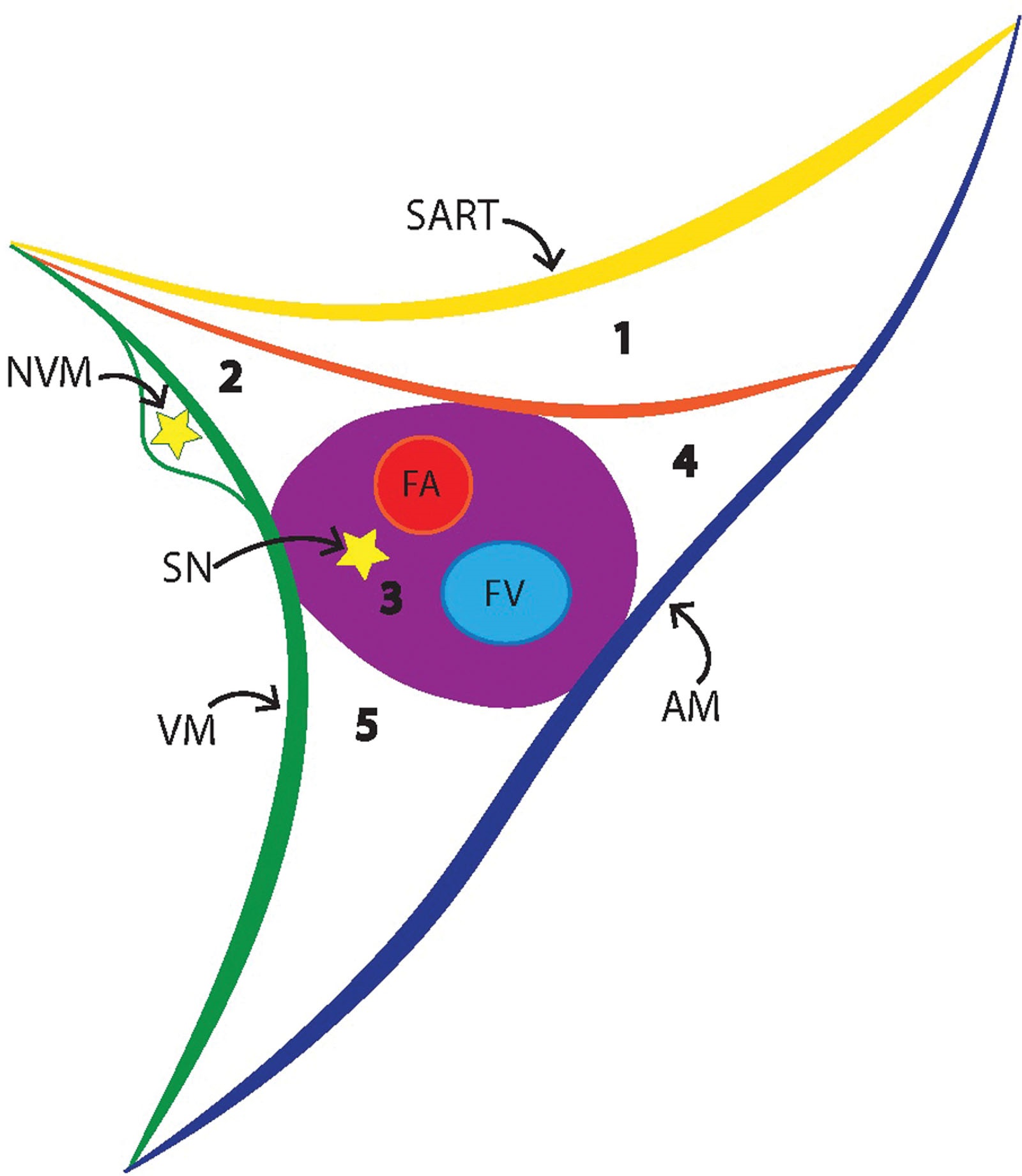
- The lateral border of the femoral triangle is the medial border of the sartorius muscle (SAM), and its medial border is the medial border of the adductor longus muscle (ALM). The apex of the femoral triangle is formed by the intersection of the medial borders of the sartorius and ALMs (View Highlight)
- The apex of the femoral triangle defines the proximal border of the adductor canal (View Highlight)
- A key anatomical feature of the distal femoral triangle and adductor canal is the presence of fascia that extends from the fascia of the vastus medialis to the fascia of the adductor muscles, effectively forming a distinct triangular subsartorial canal that contains the FA and some of the nerves (View Highlight)
- The walls of the subsartorial canal wall are formed by the VM (anterolateral wall), ALM or magnus muscles (posteromedial wall), and a fascial roof (vastofemoral fascia in the distal femoral triangle and vastoadductor membrane [VAM] in the adductor canal) (View Highlight)
- As the SN and femoral vessels enter the adductor canal, the SN is lateral to the FA. Within the adductor canal, the SN moves to a more dorsal position, relative to the artery where it (or some of its branches) may pierce the VAM (View Highlight)
- The NVM has been described as traveling with the SN into the region of the adductor canal (even though it is in its own fascial tunnel and is separated from the femoral vessels and SN) (View Highlight)
- Some have proposed that the NVM is a more important contributor to the sensory innervation to the knee than the SN. This is due to the sensory fibers to the knee contained within the NVM and its terminal branch, the medial retinacular nerve (View Highlight)
 (View Highlight)
- local anesthetic can spread distally from the distal femoral triangle to the adductor canal (View Highlight)